What is Glaucoma?
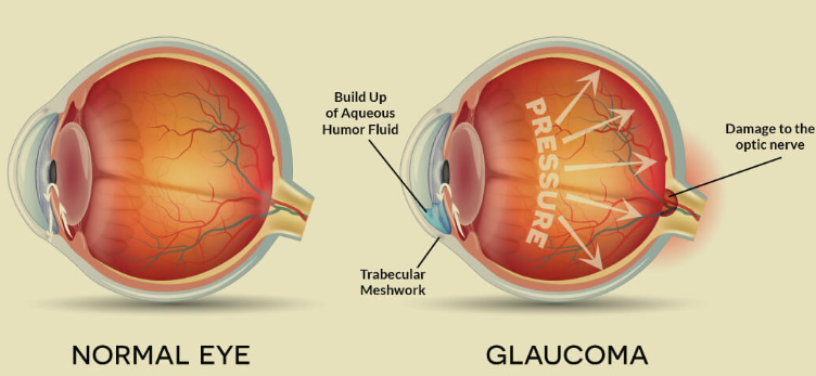
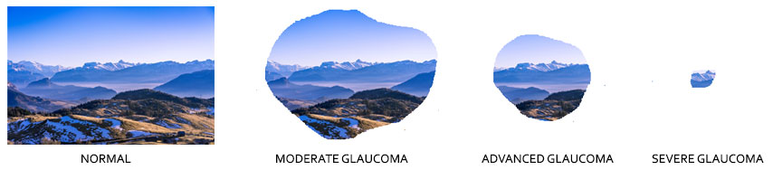
Glaucomas are a group of diseases having in common gradual loss of retinal nerve fibers leading to characteristic excavated appearance (called glaucomatous cupping) of the starting point (head) of the optic nerve (nerve responsible for carrying visual senses from eye to the brain). Neglected, this fell disease gradually devours your field of vision from the periphery and ultimately leads to complete blindness. That is why it is called the ‘silent killer of sight’ and at present, the second leading cause of blindness world-wide. High intra-ocular pressure, as opposed to our previous belief, is no longer considered an essential pre-requisite for occurrence of glaucomatous damage, though lowering eye pressure still remains the mainstay of its treatment.
What is intra-ocular pressure (IOP)?
- It is the pressure inside the eye needed to maintain the shape of the globe, as well as the normal function of the eyeball.
- It is maintained by the balance between formation and drainage of an intraocular fluid called aqueous humor, which bathes and nourishes different structures of the eye and maintains its shape. Normally the fluid drains out of the eye through a ‘drainage channel’ located at the ‘angle’ between cornea and iris.
- Obstruction to these very drainage channels causes subsequent rise of eye pressure. Although infants and children can be affected rarely, glaucoma is more common in the adult population.
Causes & types of Glaucoma
The cause of optic nerve head damage in glaucoma is not yet fully known. In most glaucoma patients, it is linked to rise in IOP which can causes lowly progressing irreversible damage to the optic nerve and can result in permanent loss of vision.
Also, patients with high IOP may never have glaucoma, and also patients with normal IOP can develop severe glaucoma.
Types of glaucoma:–
- Open angle glaucoma – the angle is structurally open but the filter beyond is blocked. Can be of two types
- Primary – Not associated with any other eye diseases
- Secondary – Associated with other eye diseases
- Angle closure glaucoma– the angle is structurally closed. Can be of two types
- Primary – Not associated with any other eye diseases
- Secondary – Associated with other eye diseases
Signs & Symptoms
- No symptoms at all. The most common. Found in open angle glaucoma. In the early stages, there is loss of peripheral vision (which patients usually do not detect by themselves, and this is the phase where we can halt the deterioration further) and in the later stages, the central vision is affected and finally the patient becomes completely and incurably blind
What pre-operative investigations are required before surgery?
At Verma Hospital, the following pre-operative investigations are advised
- Biometry – To determine the power of intraocular lens (IOL).
- Specular microscopy – To determine the health of the cornea.
- Syringing – To check the patency of the passage between the eye and nose.
- Corneal topography – To detect corneal aberrations if you are considering toric or multifocal IOLs.
- OCT (optical coherence tomography) – To detect retina abnormalities in patients with suspected retinal problems.
- USG – To detect structural (not functional) condition of retina in patients with very advanced cataracts.
- RAM – to predict the vision after surgery (not possible for very advanced cataracts).
- Blood pressure.
- ECG.
- Blood glucose (both fasting and post-prandial for diabetic patients).
- Physician clearance – For patients with other health issues (heart disease, breathing trouble, etc).
Investigations
- Tonometry – to measure eye pressure
- Gonioscopy – to assess the drainage angle
- Automated perimetry – to assess visual field damage
- Optical Coherence Tomography
Treatment
- Glaucoma cannot be cured or reversed. It can only be controlled reasonably by reducing eye pressure by –
- Medicines
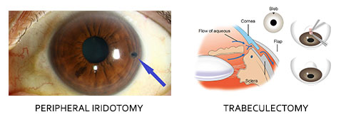
- Lasers – peripheral iridotomy – creating an alternate channel in angle closure
- Surgery – trabeculectomy, valves and shunts – by creating an alternate path in open angle as well as angle closure glaucoma
Risk factors for developing glaucoma
- Family History – parent or sibling
- Age above 40 years
- Myopia, hypermetropia
- Diabetes mellitus
- Hypertension, hypotension
- Hypothyroidism
- Eye inflammation, trauma, neglected cataract
- Long term use of steroid medicines (drops or tablets)